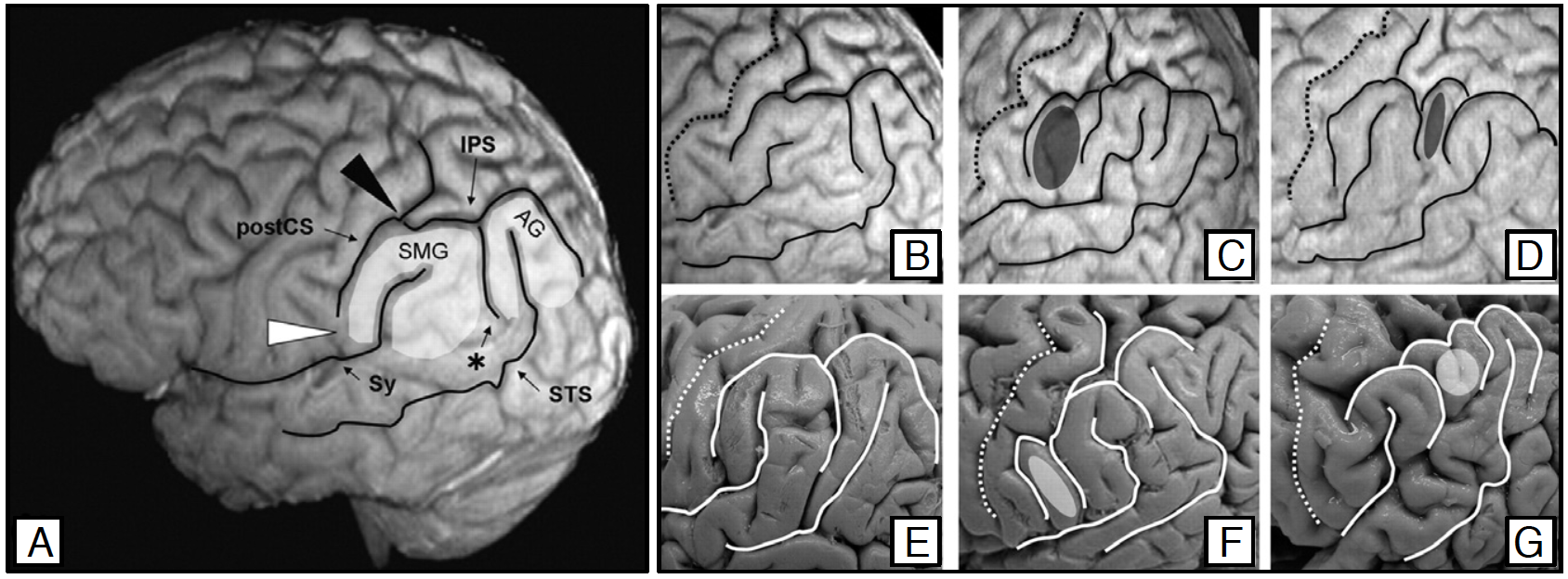
Example of natural morphological variability: left inferior parietal lobule (IPL; figure from [Kiriyama et al. 2009]). (A-D) are MRI data and (E-G) are post-mortem specimens. (A) IPL is highlighted and folds are outlined. (B,E) Typical folding pattern. (C,F) PreSMG pattern: an additional gyrus (ellipse) lies between postCS and SMG. (D,G) PreAG pattern: an additional gyrus (ellipse) lies between SMG and AG. [SMG: supramarginal gyrus; AG: angular gyrus; postCS: postcentral sulcus; IPS: intraparietal sulcus; Sy: Sylvian fissure, STS: superior temporal sulcus; *sulcus intermedius primus] postCS IPS * * Sy STS IPL

 
  Part of: Klein A (2016) Brain Graph Interface. Research Ideas and Outcomes 2: e8817. https://doi.org/10.3897/rio.2.e8817