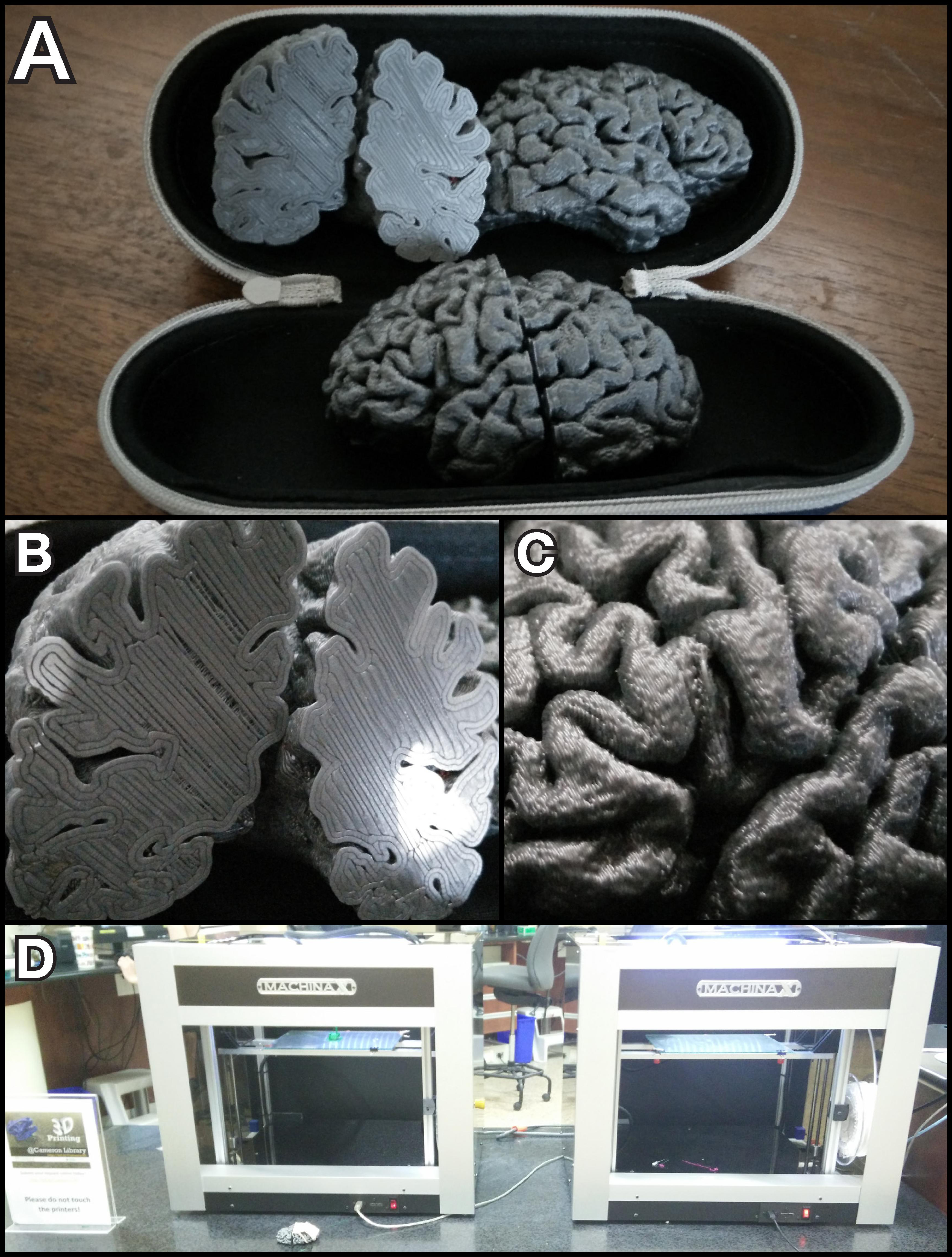
Photos of a resulting 3D-printed surface. (A) View of the full models. (B) Close-up of the coronal cross-section. (C) Close-up of the lateral surface. (D) Photo of the Machina Mk2 X20 printers used to print the models, located at the University of Alberta Libraries.